Advanced Diagnostic Procedures
Special testing is occasionally needed to diagnose or monitor an eye health problem. We constantly invest time and resources to provide you with state of the art care. The following specialized instruments may be utilized.
Age Related Macular Degeneration
 Age Related Macular Degeneration is a leading cause of vision loss and blindness in patients over the age of 60. The AdaptDx Pro allows us to detect macular degeneration in its earliest stages. Early detection and treatment are keys to preventing permanent vision loss.
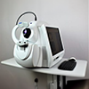
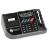
Diopsys Nova VEP and ERG
 The Diopsys Nova encompasses a technological breakthrough in eye care. Visual Evoked Potential (VEP) and Electroretinogram (ERG) testing used to only be available at a handful of research clinics. Now we can offer this advanced testing in our office. “Objective” testing means no response is necessary from the patient. All you have to do is look at a computer screen a few feet away. ERG testing has allowed us to diagnose glaucoma earlier and rule it out in questionable cases even being able to discontinue medication in some patients. VEP has allowed us to diagnose and manage patients with optic neuritis secondary to multiple sclerosis (“MS”) as well as other neurological conditions.
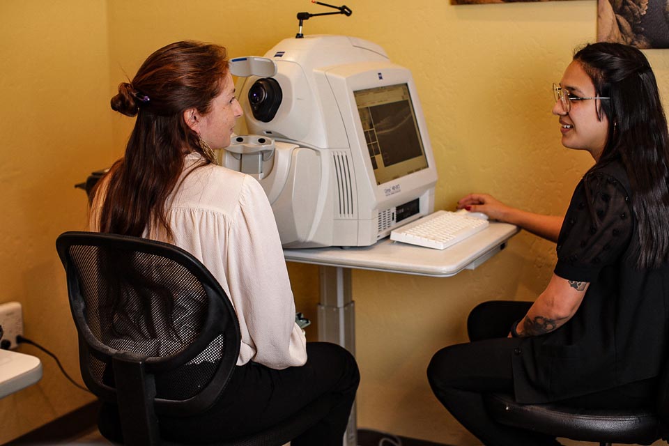
Optovue iVue SD OCT
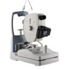
 We are so excited to add the iVue scanning laser to our practice. This Spectral Domain OCT uses 26,000 A-scans per second to give us incredible detection of the earliest signs of glaucoma, macular degeneration, and diabetic macular edema to name a few conditions. Testing is painless and takes less than two minutes. Our doctors will explain your results to you same day.
Sonogage Ultrasound Corneal Pachometer
 The Sonogage ultrasound corneal pachymeter measures the thickness of the cornea in micrometers. We use this instrument most often for glaucoma management and LASIK co-management. For glaucoma, we know from multiple studies that a thin cornea puts a patient at increased risk of glaucoma. LASIK removes corneal tissue using a femtosecond laser so corneas have to be thick enough pre-operatively to be stable after surgery.
Canon Digital Imaging System with Synemed Software
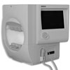
 If a picture is worth a thousand words, a high-resolution image of the eye is priceless. Our imaging system utilizes a low intensity flash to increase patient comfort. Using the Synemed software, images can be magnified, colors can be inverted, and subtle changes can be detected when comparing images to previous results. This not only helps our doctors effectively manage your condition but assists in your understanding as well.
Humphrey Visual Field Analyzer
 Humphrey visual fields are the gold standard in eye care. Visual field defects are noted in glaucoma, strokes, macular degeneration and many more conditions. Every comprehensive eye exam includes a screening visual field at no additional charge. If further study is needed, our doctors will order a threshold visual field test. This testing parameter detects more subtle visual field defects and allows close monitoring over time.
Diagnostic Sets of Intracanalicular Plugs For Dry Eye Therapy
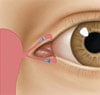 Dry Eye Syndrome is very common in northern Arizona. After a full diagnostic work up for Dry Eye Syndrome, our doctors may recommend intracanalicular plugs as part of the treatment regimen. Diagnostic plugs allow you to experience a 4-7 day test period to determine if you may benefit from longer lasting plugs. After the test period, the plugs will simply dissolve on their own. If you experience temporary improvement of your symptoms, our doctors will discuss longer lasting options to achieve ongoing relief. Read more about intracanalicular plugs
Topcon Corneal Topographer
 Our corneal topographer provides a three-dimensional view of the front surface of the eye (cornea). This testing is utilized in diagnosing and monitoring keratoconus, epithelial-basement-membrane-dystrophy, irregular forms of astigmatism, and in fitting certain kinds of contact lenses.